Areas of Specialty
Shoulder:
The shoulder’s amazing 180-degree range of motion on three different planes makes it a remarkable joint. However, because the more a joint can do, the more can go wrong. Specialized treatment is a must for a complex body part like the shoulder. Klamath Orthopedic physicians have the experience to provide comprehensive care of the shoulder including non-surgical treatments, arthroscopic, and open surgeries.
Elbow:
The elbow is more than a hinge. The complex arrangement of muscles, ligaments and tendons that allow for rotation help us perform daily functions – including fun, competitive activities such as sports. Treatment of injuries and diseases of the elbow requires specialized knowledge. Our physicians are experienced in the diagnosis and treatment of elbow problems. They provide an extraordinary base of shared knowledge with leading-edge surgery and treatment.
Conditions we treat include:
-
- Arthritis of the Shoulder/Elbow
- Shoulder and Elbow Fractures
- Shoulder Dislocation
- Overuse Elbow Injuries (ex: Tennis Elbow)
- Shoulder and Elbow Sprains
- Shoulder Labral Tear
- Shoulder Tendinitis
- Rotator Cuff Tears
- Shoulder Instability
Surgeries include:
-
- Total Shoulder Replacement
- Reverse Total Shoulder Replacement
- Arthroscopic Rotator Cuff Repair
- Arthroscopic Labral Repair
- Arthroscopic Elbow Surgery
Spine
When you have neck, spine or back pain; 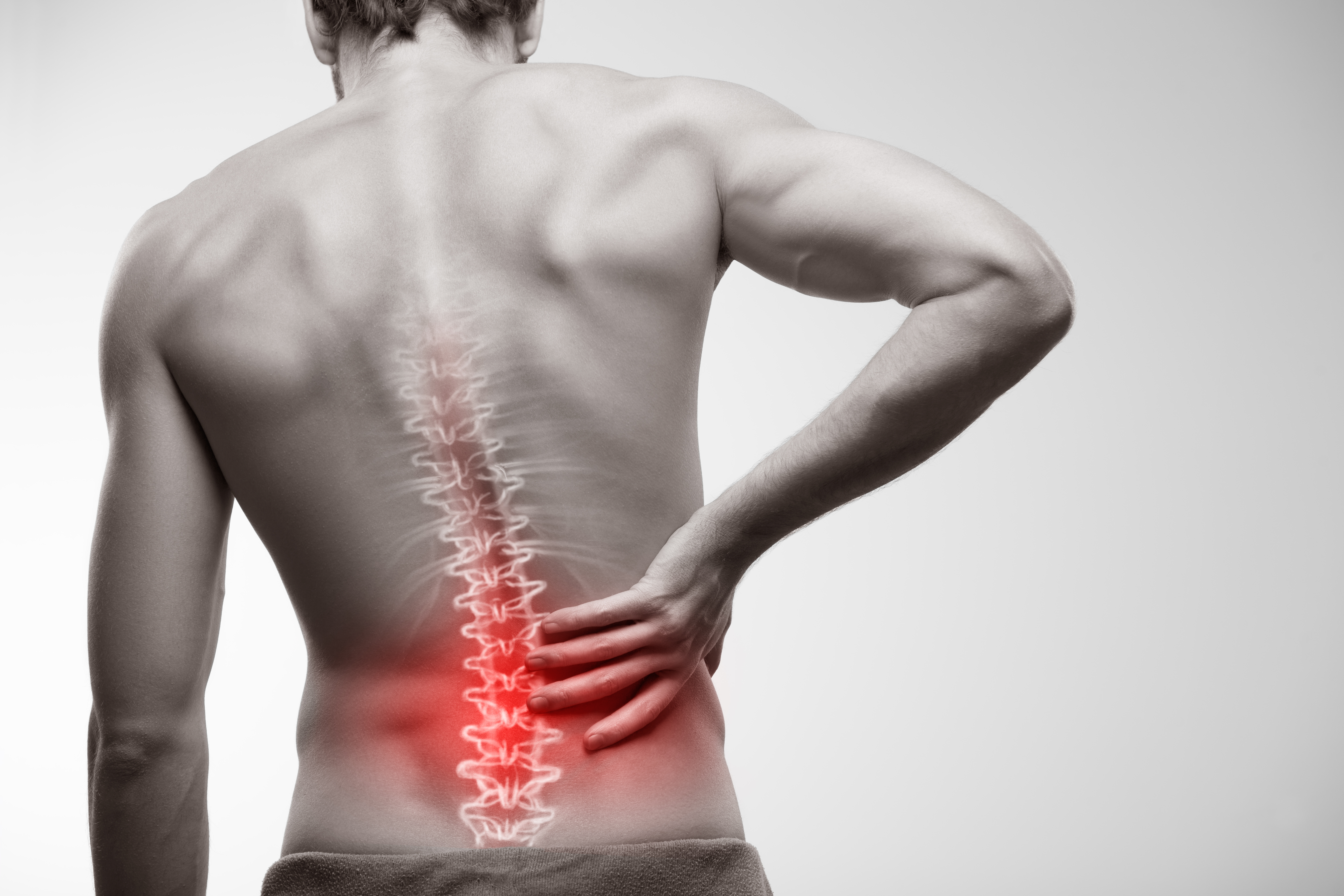 you don’t want to wait around and hope things get better – neither do we. Our number-one goal is to get you back to your daily activities. The medical treatment required for spinal injuries and diseases requires advanced knowledge because the spine is one of the most complex and important systems in the human body. Klamath Orthopedic’s spine specialists focus on restoration of function, reduction in pain, and control of costs in developing any treatment for back pain. If non-surgical approaches are not effective, the team is able to offer the latest advancements in state-of-the-art spine surgery.
Conditions We Treat Include
Cervical Procedures
-
- Posterior Cervical Decompression
- Posterior Cervical Fusion
- Anterior Cervical Decompression with Fusion
- Cervical Artificial Disc Replacement
Lumbar Procedures
-
- Laminotomy with Posterior Element Decompression
- Transforaminal Lumbar Interbody Fusion (TLIF)
- Posterior Lumbar Interbody Fusion (PLIF)
- Microdiscectomy
- Lumbar Discectomy and Neural Decompression
- Foraminotomy
- Laminotomy Decompression for Bony Stenosis
- Extreme Lateral Interbody Fusion (XLIF)
- Lumbar Artificial Disc Replacement
Spinal Conditions Treated
-
- Congenital spinal malformations
- Disc herniation
- Radiculopathy & Myelopathy
- Scoliosis
- Spinal fractures
- Spinal stenosis (Narrowing)
- Spondylolisthesis (Vertebrae “slipping”)
- Osteoporosis
- Disc degeneration
- Spondylosis (Arthritis of the spine)
Surgeries Include:
-
- Discectomy
- Fusion
- Laminectomy
- Laminotomy
Hip
 Your hips are a part of your daily life – sitting, standing, laying and bending. The cup-and-ball-like joint allows for a wide range of motions. The medical care for hips has changed sigificantly in recent years as technology and procedures become less invasive and more advanced. The surgeons at Klamath Orthopedic provide this state-of-the-art care for hips through minimally invasive surgical intervention and full replacements. Our physicians treat individuals based on their needs with the right approach at the right time for every patient.
Conditions we treat include:
-
- Trochanteric Bursitis
- Labral Tears
- Synovitis
- Cartilage Injury
- Femoroacetablar Impingement (FAI)
- Abductor Tendon Tears
- Sprains and Strains
- Overuse Injuries
- Arthritis
- Snapping Hip Syndrome
Surgeries include:
-
- Hip Replacement
- Arthroscopic Snapping Hip Releases
- Arthroscopic Bursectomy
.
Hand and Wrist
The hand and wrist are intricate, containing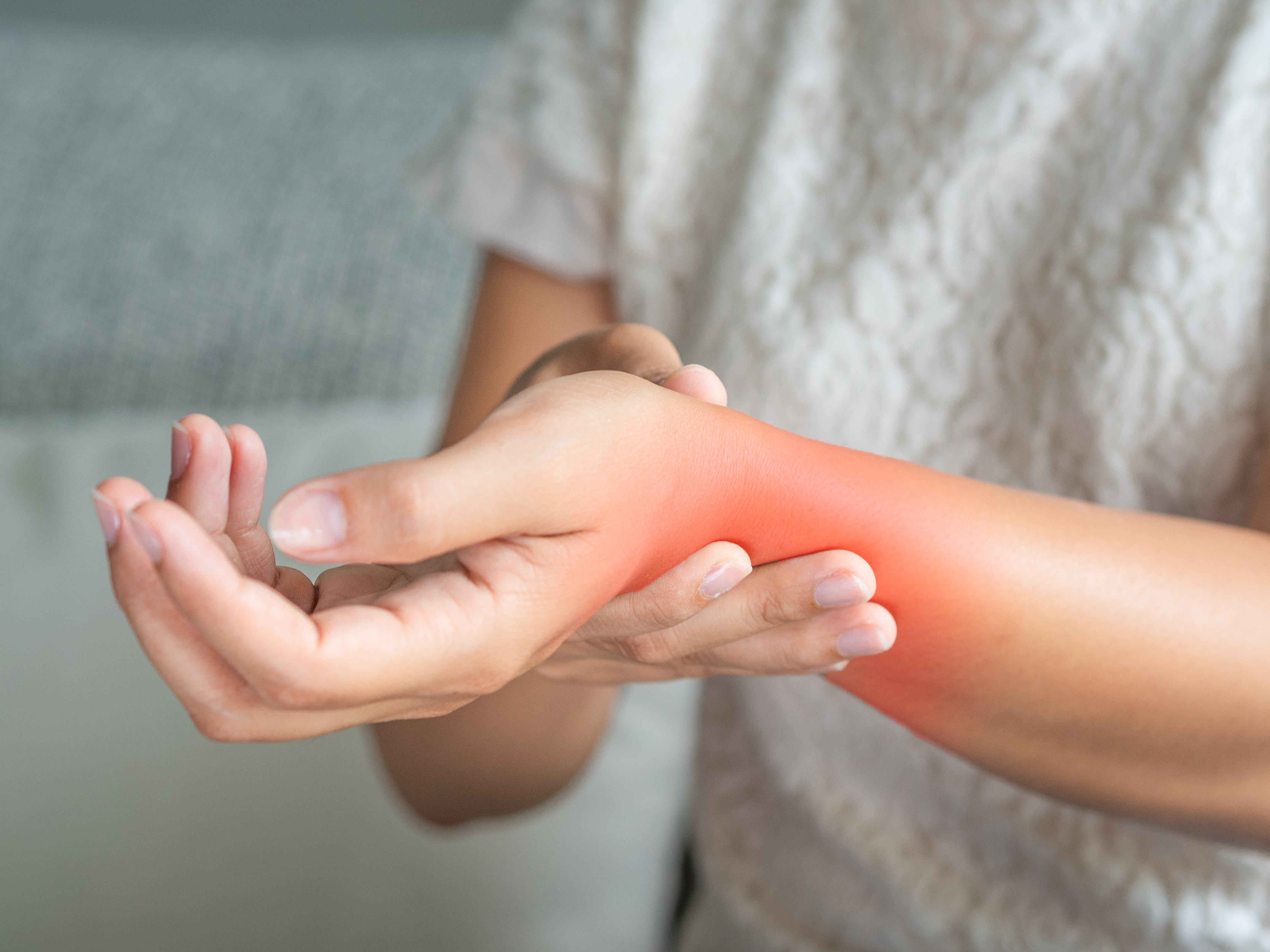 dozens of bones, joints, ligaments, tendons and muscles that need to work together seamlessly every day. Because they are so essential to daily living, they require highly-specialized medical care when they become injured. Klamath Orthopedic offers expertise in conditions of the hand, wrist and upper extremities. Our goal is to treat with the least invasive approach first, and follow with surgery only if necessary. Our physicians specialize in minimally-invasive arthroscopic surgical techniques, as well as endoscopic carpal and cubital tunnel release that reduce pain and quicken recovery.
Conditions We Treat
-
- Carpal tunnel syndrome, cubital tunnel syndrome, radial tunnel syndrome
- Fractures and dislocations of wrist, hand, distal radius, and distal ulna
- Arthritis of the thumb, hand, or wrist
- Sports-related injuries of the hand or wrist
- Trigger finger
- Tendonitis of hand, wrist, or elbow
- Tendon, nerve, and blood vessel injuries
- Vascular disease or circulatory disturbances in the hand
- Work-related injuries and conditions
Surgeries Performed
-
- Carpal tunnel release
- Dupuytren’s contracture fasciectomy
- Trigger finger release
- Tendon Repair
- Ganglion removal
Knee Repair
The knee is one of the largest and most complex joints in the body. The knee joins the thigh bone (femur) to the shin bone (tibia). The smaller bone that runs alongside the tibia (fibula) and the kneecap (patella) are the other bones that make the knee joint.
Tendons connect the knee bones to the leg muscles that move the knee joint. Ligaments join the knee bones and provide stability to the knee:
- The anterior cruciate ligament prevents the femur from sliding backward on the tibia (or the tibia sliding forward on the femur).
- The posterior cruciate ligament prevents the femur from sliding forward on the tibia (or the tibia from sliding backward on the femur).
- The medial and lateral collateral ligaments prevent the femur from sliding side to side.
Two C-shaped pieces of cartilage called the medial and lateral menisci act as shock absorbers between the femur and tibia.
Numerous bursae, or fluid-filled sacs, help the knee move smoothly.
Conditions We Treat:
- Chondromalacia patella (also called patellofemoral syndrome): Irritation of the cartilage on the underside of the kneecap (patella), causing knee pain. This is a common cause of knee pain in young people.
- Knee osteoarthritis: Osteoarthritis is the most common form of arthritis, and often affects the knees. Caused by aging and wear and tear of cartilage, osteoarthritis symptoms may include knee pain, stiffness, and swelling.
- Knee effusion: Fluid build-up inside the knee, usually from inflammation. Any form of arthritis or injury may cause a knee effusion.
- Meniscal tear: Damage to a meniscus, the cartilage that cushions the knee, often occurs with twisting the knee. Large tears may cause the knee to lock.
- ACL (anterior cruciate ligament) strain or tear: The ACL is responsible for a large part of the knee’s stability. An ACL tear often leads to the knee “giving out,” and may require surgical repair.
- PCL (posterior cruciate ligament) strain or tear: PCL tears can cause pain, swelling, and knee instability. These injuries are less common than ACL tears, and physical therapy (rather than surgery) is usually the best option.
- MCL (medial collateral ligament) strain or tear: This injury may cause pain and possible instability to the inner side of the knee.
- Patellar subluxation: The kneecap slides abnormally or dislocates along the thigh bone during activity. Knee pain around the kneecap results.
- Patellar tendonitis: Inflammation of the tendon connecting the kneecap (patella) to the shin bone. This occurs mostly in athletes from repeated jumping.
- Knee bursitis: Pain, swelling, and warmth in any of the bursae of the knee. Bursitis often occurs from overuse or injury.
- Baker’s cyst: Collection of fluid in the back of the knee. Baker’s cysts usually develop from a persistent effusion as in conditions such as arthritis.
- Rheumatoid arthritis: An autoimmune condition that can cause arthritis in any joint, including the knees. If untreated, rheumatoid arthritis can cause permanent joint damage.
- Gout: A form of arthritis caused by buildup of uric acid crystals in a joint. The knees may be affected, causing episodes of severe pain and swelling.
- Pseudogout: A form of arthritis similar to gout, caused by calcium pyrophosphate crystals depositing in the knee or other joints.
- Septic arthritis: Bacterial infection inside the knee can cause inflammation, pain, swelling, and difficulty moving the knee. Although uncommon, septic arthritis is a serious condition that usually gets worse quickly without treatment.
Foot and Ankle
 The foot is made up of 26 bones, 33 joints, 107 ligaments, 19 muscles, and numerous tendons. Complex biomechanics keep all these parts in the right position and moving together. Given these intricacies, it is not surprising that most people will experience some problem with their feet at some time in their lives. Foot pain can be detrimental to continuing an active, healthy lifestyle. At Klamath Orthopedic, we focus on the diagnosis and treatment of conditions that affect the foot and ankle, promoting long-term health.
Conditions
• Ankle pain
• Ankle sprain
• Ankle arthritis
• Bunions
• Congenital ankle and foot deformities
• Diabetic limb
• Drop Foot
• Flat feet & high arch feet
• Fractures
• Gout
• Hammertoes
• Heel pain
• Infections (Bacterial, Fungal, Viral)
• Ingrown nails
• Nerve problems & neuromas
• Skin Conditions
• Sports injuries
• Tendonitis
• Tendon tears
• Tumors
• Achilles Tendon Rupture
• Ankle Fractures (Broken Ankle)
• Calcaneus (Heel Bone) Fractures
• Dislocations
• Jones Fracture (5th metatarsal base)
• Lisfranc (Midfoot) Injury
• Pilon Fractures of the Ankle
• Stress Fractures of the Foot and Ankle
• Talus Fractures Toe and Forefoot Fractures
• Turf Toe
Surgeries
All of our physicians are board certified by the American Board of Foot and Ankle Surgery. Our clinic offers specialized care in all aspects of foot and ankle surgery including reconstructive surgery for common and complex deformities, arthritis, trauma surgery, and arthroscopic (minimally invasive) surgery.
Surgical treatments
- Total Ankle Replacement
- Ankle ligament reconstruction for chronic ankle sprain
- Flatfoot reconstructions & realignments
- Bunion, hallux rigidus and forefoot reconstruction
- Surgery for foot and ankle fractures
- Ankle fusion or ankle replacement for arthritis
- Diagnostic ankle arthroscopy
- Arthroscopic ankle cartilage repair
- Lateral ankle ligament reconstruction
- Surgery for foot deformities
Nonsurgical treatments
- Exercise
- Injections
- Medication
- Orthotics
- Physical therapy
- Splinting
Ankle
- Talocrural Joint: This is a hinge joint formed by the distal ends of the fibula and tibula that enclose the upper surface of the talus. It allows for both dorsiflexion (decreasing the angle between the foot and the shin) and plantarflexion (increasing the angle).
- Inferior tibiofibular Joint: This is strong joint between the lower surfaces of the tibia and fibula. This is supported by the inferior tibiofibular ligament.
- Subtalar Joint: This joint comprises of the articulating surfaces of the talus and the calcaneus. It provides shock absorption and the movements of inversion and eversion (inward and outward ankle movements respectively) occur here.
Ligaments of the ankle joint
The ligaments of the ankle joint are comprised mainly of the collateral ligaments, both medial (inner) and lateral (outer). These are extremely important in the stability of the ankle itself:
- Lateral Collateral Ligament:
The lateral collateral ligament prevents excessive inversion. It is considerably weaker than the larger medial ligament and thus sprains to the lateral ligament are much more common. It is made up of 3 individual bands:
- Anterior talofibular ligament (AFTL): passes from the fibula to the front of the talus bone.
- Calcaneofibular ligament (CFL)– connects the calcaneus and the fibula
- Posterior talofibular Ligament (PTFL)– passes from the back of the fibula to the rear surface of the calcaneus.
- Medial Collateral Ligament:
The medial ligament also known as the deltoid ligament is considerably thicker than the lateral ligament and spreads out in a fan shape to cover the distal (bottom) end of the tibia and the inner surfaces of the talus, navicular, and calcaneus.


